Reflectivity 3.2
Update: Reflectivity is now commercialized by Abyss Processing. It was fully revamped and offers 3D AI solutions for glaucoma. Please visit the Abyss Processing website to request a trial license.
What is Reflectivity 3.2?
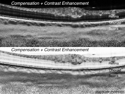
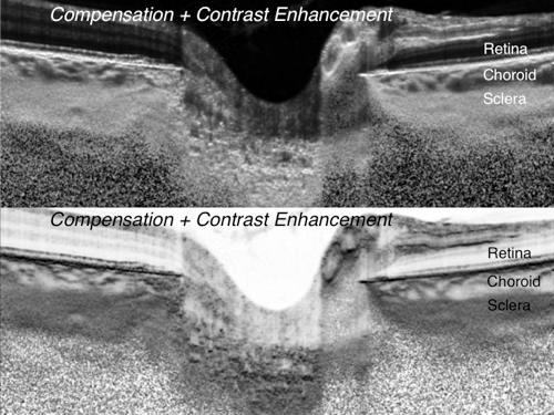
Reflectivity is a post-processing software that can improve the quality of medical images captured with Optical Coherence Tomography (OCT), Ultrasounds, or Phtoacoustic Imaging.
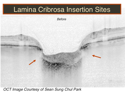
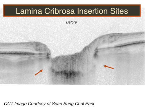
For Optic Nerve Head (ONH) images captured with OCT, Reflectivity can:
- Remove blood vessel shadows in the ONH tissues
- Improve the visibility of the anterior lamina cribrosa
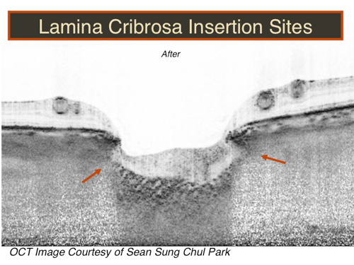
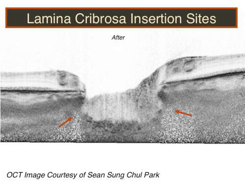
- Improve the visibility of lamina/sclera insertions
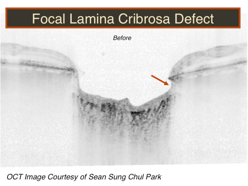
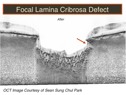
- Improve the visibility of lamina cribrosa defects
- Improve the visibility of Drusen regions
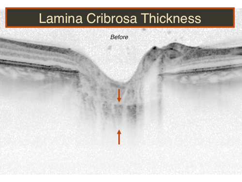
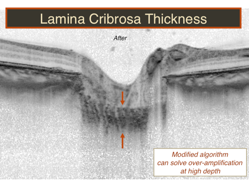
- Help identify the full thickness of the lamina cribrosa
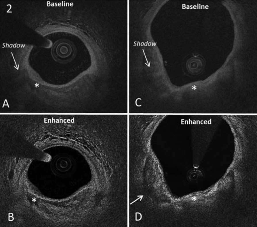
For blood vessel images captured with intravascular OCT, Reflectivity can:
- Enhance plaque and blood vessel layer contrast
- Remove shadow artefacts from dense structures
- Improve the visibility of the deepest tissue layers
- Allow better identification of external plaque boundaries
Please note that not all the above can be achieved in every sample so care must be taken when interpreting the data.
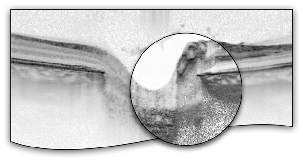
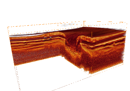
Examples
Original Images
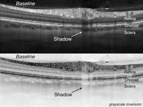
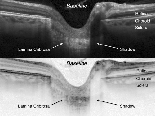
Enhanced Images
To Learn More About Reflectivity
For more information, please see:
- Girard MJA, Strouthidis NG, Ethier CR, Mari JM. Shadow Removal and Contrast Enhancement in Optical Coherence Tomography Images of the Human Optic Nerve Head. Invest Opthalmol and Vis Sci. 2011; 52(10):7738-48. Pdf. Pubmed. IOVS.
- Foin N, Mari JM, Davies JE, Di Mario C, Girard MJA. Imaging of Coronary Artery Plaques using Contrast-enhanced Optical Coherence Tomography. European Heart Journal – Cardiovascular Imaging. 2013; 14(1):85. Pdf. Pubmed.
- Mari JM, Park SC, Strouthids NG, Girard MJA. Enhancement of Lamina Cribrosa Visibility in Optical Coherence Tomography Images Using Adaptive Compensation. Invest Ophthalm and Vis Sci. 2013; 54(3):2238-47. Pdf. Pubmed. IOVS.
- Foin N, Mari JM, Di Mario C, Davies JE, Girard MJA. Intracoronary Imaging using Attenuation-compensated Optical Coherence Tomography Allows Better Visualisation of Coronary Artery Diseases. Cardiovascular Revascularization Medicine. 2013; 14(3):139-143. Pdf. Pubmed.
- Kim TW, Wollstein G, Leung C, Girard MJA, Strouthidis NG, Sung KR, Kagemann L, Schuman J. Imaging of lamina cribrosa in glaucoma: Perspectives of pathogenesis and clinical applications. Current Eye Research. 2013; 38(9):903-9. Pdf. Pubmed.
Or contact us at:
mgirard@ophthalmic.engineering
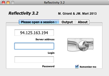
Instructions for Reflectivity 3.2
Reflectivity 3.2 is a user-friendly software that can run on both Mac and Windows Platforms. A collaborative agreement with our laboratories needs to be reached before one can use reflectivity.
To use Reflectivity 3.2, the user needs an internet connection and a software account (User name and Password). To obtain your account, please contact us. The software account will be locked to your computer, so if you need to use Reflectivity on a different computer, you will need an additional account.
Reflectivity 3.2 will let you compensate OCT and Ultrasound image data not only in the .vol format (from Heidelberg Engineering) but now also in .png, .jpg, .gif, .bmp, and .tif formats. Two control parameters (decompression and compression exponents) can be used to optimize the compensation. We recommend that you start with the default values provided. In brief, compensation can now be applied to data gathered with other OCT devices (Cirrus, Bioptigen, etc) or Ultrasound devices.
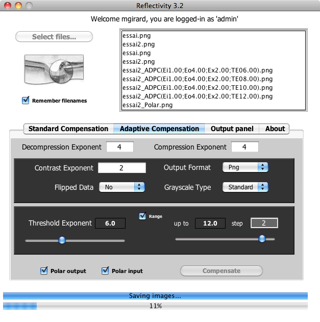
Reflectivity 3.2 will let you compensate several files in batch mode. For example, when you click 'select files', just hold SHIFT to select all the images/volumes you want to compensate. Compensation will be performed on all selected files. This will considerably save time for the user if many files need to be processed.
Reflectivity 3.2 allows the user to perform either standard compensation or adaptive compensation.
Standard Compensation
Standard compensation was the first algorithm that was released to improve the quality of OCT images. The user can define the contrast exponent to improve the visibility of tissue boundary. A contrast exponent of 1 indicates that no contrast enhancement is performed. We recommend a value of 2, as higher values can generate more noise but may be useful to identify specific boundaries such as the retinal pigment epithelium. While standard compensation can provide drastic enhancement, it is limited by the presence of noise over-amplification at high depth. Noise over-amplification could limit a proper identification of the deepest tissue structures such as the lamina cribrosa in ophthalmic applications.
Adaptive Compensation
Adaptive compensation belongs to the 2nd generation of compensation algorithms. Adaptive compensation was developed to overcome the over-amplification of noise at high depth. In adaptive compensation, standard compensation operations are performed until an energy threshold is reached, at which stage the compensation process is stopped to limit noise over-amplification in the deeper portion of the OCT image. If the exponent is large, adaptive compensation will behave like the standard compensation algorithm. The smaller the threshold exponent is, the more noise will be filtered out. Care must taken not to use a too large exponent as this could impact the visual interpretation of the sturctures present within the OCT/Ultrasound images. We recommend the user to try several exponents to achieve the desired visibility before making any conclusions. A range of threshold exponents can be used to facilitate data interpretation.
Output
Reflectivity 3.2 will output both Baseline and Improved images in the format specified by the user. Files will be found in a folder of the same name as that of the input file. Reflectivity 3.2 should be straightforward to use but please let us know if you encounter any issues.
Notes on Intravascular Data
To process RAW intravascular data, ensure that the decompression exponent is set to 1. If you select the options 'polar input' and 'polar output', Reflectivity 3.2 will reconstruct the images in polar coordinates for the original image and the compensated image, respectively.
Disclaimer
Reflectivity 3.2 is provided 'as is' with no warranty of any kind. Reflectivity 3.2 has not yet been approved in a clinical setting and should not be used for making clinical decisions. Reflectivity 3.2 requires an online connection but only for the purpose of logging. Once a user is connected, Reflectivity 3.2 will run even without an internet connection. No medical data from the user are therefore uploaded onto our servers.
