Optic Nerve Head Deformations During Eye Movement
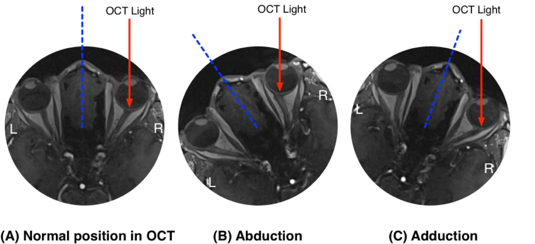
Visualization of optic nerve head deformations using OCT (Subject 1)
Visualization of optic nerve head deformations using OCT (Subject 2)
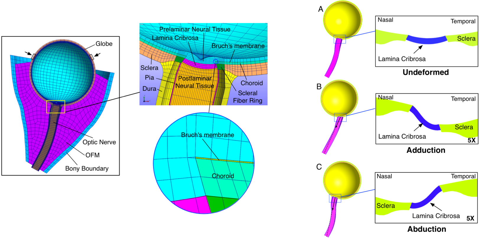
Finite element predictions
We combined finite element (FE) analysis and dynamic magnetic resonance imaging (MRI) to estimate optic nerve head (ONH) strains (deformations) during eye movements, and identify factors influencing such strains.
The eyes and orbital tissues of a healthy subject were visualized during visually-guided horizontal eye movements, using dynamic MRI. A baseline FE model of the left eye was reconstructed in the primary gaze position and included details from the orbital tissues.
The baseline FE model matched the MRI scan well. Our models predicted that, during eye movements, the retrobulbar portion of optic nerve had a pulling action on the ONH, which resulted in large strains within the ONH tissues.
In vivo measurements using OCT
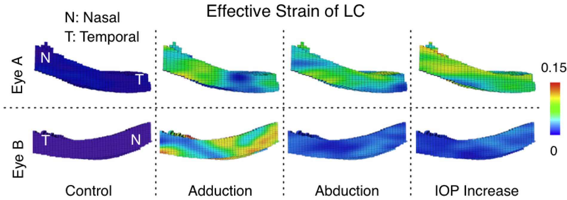
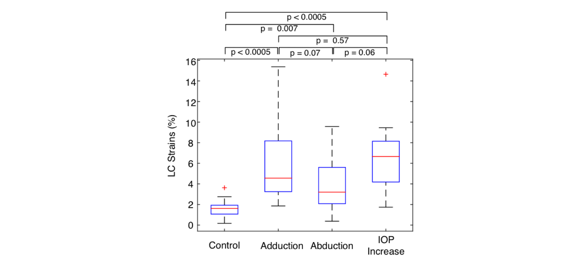
We further measured lamina cribrosa (LC) strains (deformations) following abduction and adduction in healthy subjects and to compare them with those resulting from a relatively high acute intraocular pressure (IOP) elevation using in vivo OCT imaging.
A total of 16 eyes from 8 healthy subjects were included. For each subject, both optic nerve heads (ONHs) were imaged using optical coherence tomography (OCT) at baseline (twice), in different gaze positions (adduction and abduction of 20°) and following an acute IOP elevation of approximately 20 mm Hg from baseline (via ophthalmodynamometry). Strains of LC for all loading scenarios were mapped using a three-dimensional tracking algorithm.
Our results confirm that horizontal eye movements generate significant ONH strains, which is consistent with previous estimations using finite element analysis.
The two videos above show the deformations of lamina cribrosa under different loading scenarios.